(Dr Nicola Rogers Simpson)

I am currently a Senior Research Fellow, and work with some great chemists at the University of Warwick with Professor Peter Scott. We make new molecules that wrap around iron(II) metal centres to form quite exquisite structures, a little bit like small peptides and proteins.
We work with biologists, who have discovered that some of our ‘metallohelices’ can kill bacteria such as E. coli. This is important because many bacteria are becoming resistant to the current antibiotics that we use in the clinic, and we need to desperately find new medicines that can kill bacteria.

Most of the research that I have done so far has involved looking at how the chemistry of metal ions can be exploited to develop either therapeutic or diagnostic agents for medical applications, i.e. making molecules that we can see by MRI or by fluorescence microscopy, or than can kill bacteria or cancer cells. I have worked in research for nearly 6 years following my PhD, but I also spend quite a lot of my time cycling, running, and playing the clarinet. I have worked alongside Zoe Schnepp and Ruth Patchett on the ChemBam website, developing the experiments, and putting it all together.
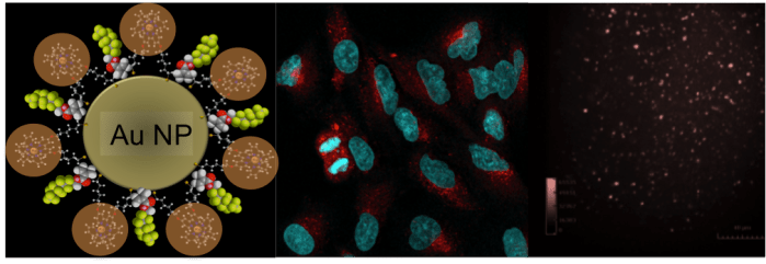
I studied Chemistry as an undergraduate at the University of Birmingham, and went on into PhD research with Professor Zoe Pikramenou at the University. During my PhD I made gold nanoparticles, and coated them in luminescent molecules, to make bright luminescent nanoparticles for imaging in cells, and in flow.
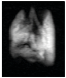
In 2014 I took on a short research position at the Sir Peter Mansfield Imaging Centre in Nottingham, and worked in the pulmonary imaging group of Professor Thomas Meersmann. I helped to develop new methods of using noble gases to image the lungs by MRI, by creating a ‘magnetic gas’ to inflate the lungs and look at lung disease.
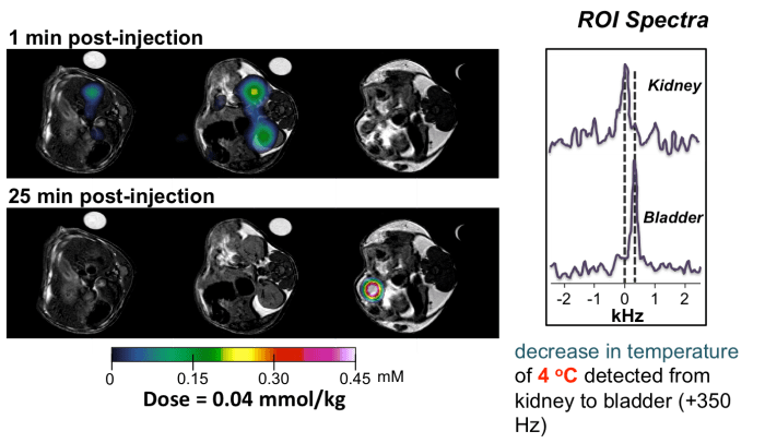
Following this MRI work I joined the group of Professor David Parker in Durham, and spent two years developing molecules for detection by MRI. We developed molecular probes, using lanthanide complexes, that are more sensitive than current contrast agents used in the clinic, and which can also act as ‘smart’ molecules to read out information in the body, such as tissue acidity and temperature.
Access my researchgate profile here.

This work is licensed under a Creative Commons Attribution 4.0 International License.
