Page created by Dr Maria Chiara Arno
Sir George Stokes, a British scientist, first discovered fluorescence in 1852 when he observed that the mineral fluorite (Fig. 1, molecular formula CaF2) emitted red light when it was illuminated by ultraviolet excitation. Early investigations in the 19th century showed that many specimens (including minerals, crystals, drugs, butter, chlorophyll, and vitamins) fluoresce when irradiated with ultraviolet light.
Fig. 1. Fluorite, fluorescent under UV light
Fluorescence is the temporary absorption of electromagnetic wavelengths from the visible light spectrum by fluorescent molecules, and the subsequent emission of light at a lower energy level.
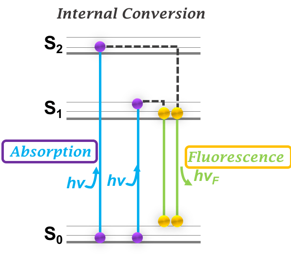
Stimulating light excites an electron, raising energy to an unstable level. This instability is unfavourable, so the energized electron is returned to a stable state almost as immediately as it becomes unstable. This return to stability corresponds with the release of excess energy in the form of fluorescence light (Fig. 2).
Fig. 2. Jablonski diagram. After an electron absorbs a photon the system is excited electronically and vibrationally. When the system relaxes vibrationally, it fluoresces at a longer wavelength. Figure author: Yujie Xie.
In general, emitted fluorescence light has a longer wavelength and lower energy than the absorbed light. This phenomenon, known as Stokes shift, can be ascribed to energy loss between the time a photon is absorbed and when a new one is emitted.
It was not until the 1930s that the use of fluorophores was initiated in biological investigations to stain human tissues, bacteria, and viruses. Several of these stains were highly specific and stimulated the development of the first fluorescence microscope.
Fluorescence microscopy in biology
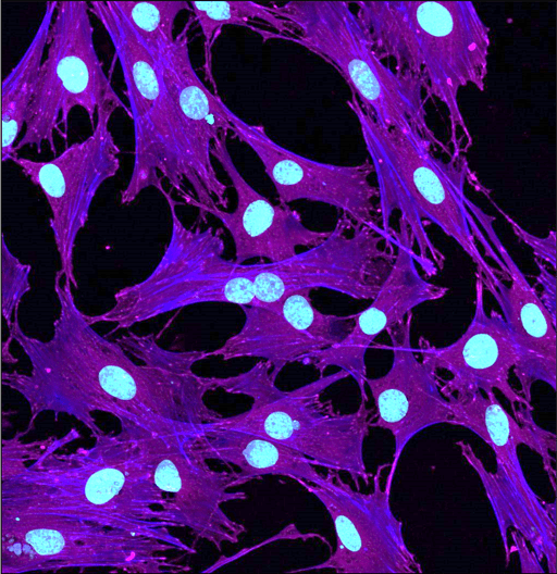
Fig. 3. Human cells stained in cyan at the nuclei and purple-blue at the cytoskeleton
The technique of fluorescence microscopy has become an essential tool in biology and the biomedical sciences, as well as in materials science. The application of an array of fluorophores has made it possible to identify cells and sub-microscopic cellular components with a high degree of specificity (Fig. 3). These molecules can be conjugated to the regions of interest, such as cellular matrices or sub-cellular organelles and are excited by specific wavelengths. Through the use of multiple fluorescence labels, different probes can be used to identify several targets simultaneously.
In materials science, fluorescence microscopy can be used to identify the shape and size of nanoparticles, after labelling them with a fluorophore.
How does a fluorescence microscope work?
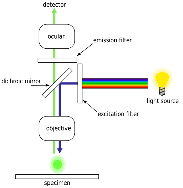
The majority of fluorescence microscopes have the design shown in the diagram on Fig. 4. Light of the excitation wavelength illuminates the specimen through the objective lens.
Fig. 4. Diagram of a fluorescence microscope design.
The fluorescence emitted by the specimen is focused to the detector by the same objective that is used for the excitation. Since most of the excitation light is transmitted through the specimen, only reflected excitatory light reaches the objective together with the emitted light, therefore giving a high signal-to-noise ratio. The dichroic mirror acts as a wavelength specific filter, transmitting fluoresced light through to the eyepiece or detector, but reflecting any remaining excitation light back towards the source.
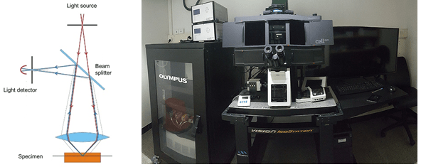
An advanced type of fluorescence microscope is the confocal microscope (Fig. 5), where a pinhole is placed in front of the illumination source to allow transmission only through a small area. This illumination pinhole is imaged onto the focal plane of the specimen, meaning only one point of the specimen is illuminated at a time. As a consequence of this, only fluorescence excited within the focal plane of the specimen will go through the detector pinhole. This will allow to achieve high signal to noise ratio (and hence better image quality) by scanning small sections at a time and then stitch them together for an overall view.
Fig. 5. Diagram of a confocal microscope design and photo of a confocal microscope.
This work is licensed under a Creative Commons Attribution 4.0 International License.